
+91-11-41543333
Category :
Cornea
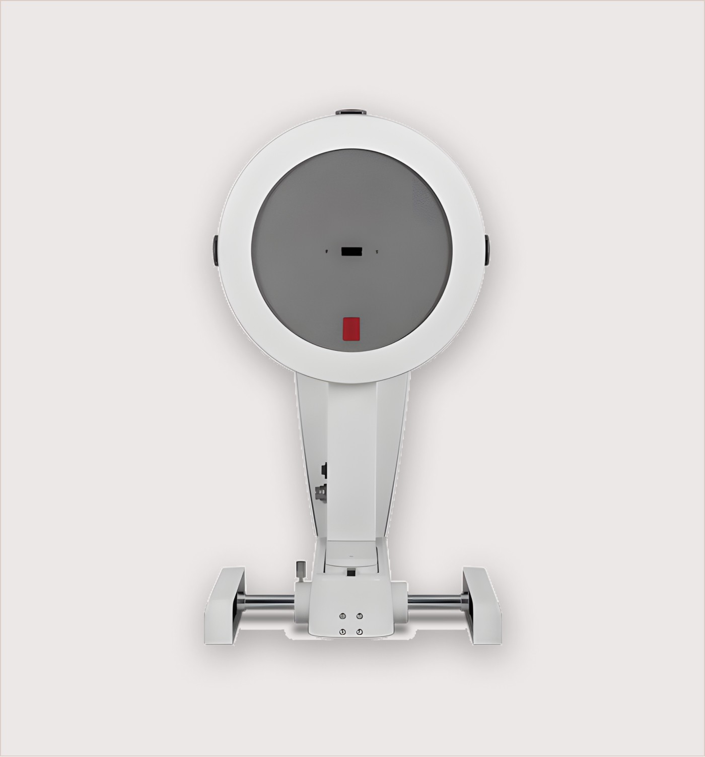
Pentacam
Pentacam
Features
Pentacam measures the cornea from limbus to limbus. It measures up to 25 ,000 true elevation points. During the rotating scan that takes max. 2 seconds, up to 50 Scheimpflug images of the anterior eye segment are captured with outstanding precision. Based on these images the Pentacam® provides topographic data on elevation and curvature of the entire anterior and posterior corneal surfaces. The corneal thickness (pachymetry) is measured and presented graphically over its entire surface. A topography based keratoconus detection and quantification are performed. The anterior chamber depth, chamber volume (size) and the chamber angles are automatically calculated and presented for glaucoma screening. The illumination of the eye using blue LED light makes corneal and lens opacities (cataract) visible. The anterior chamber can be visualised and displayed with the virtual tomography model
Contact Us for further information
Contact Us
© 2023 KLB. All rights reserved.
Contact Us
© 2023 KLB. All rights reserved.
Contact Us
© 2023 KLB. All rights reserved.
Category :
Cornea
Pentacam
Features
Pentacam measures the cornea from limbus to limbus. It measures up to 25 ,000 true elevation points. During the rotating scan that takes max. 2 seconds, up to 50 Scheimpflug images of the anterior eye segment are captured with outstanding precision. Based on these images the Pentacam® provides topographic data on elevation and curvature of the entire anterior and posterior corneal surfaces. The corneal thickness (pachymetry) is measured and presented graphically over its entire surface. A topography based keratoconus detection and quantification are performed. The anterior chamber depth, chamber volume (size) and the chamber angles are automatically calculated and presented for glaucoma screening. The illumination of the eye using blue LED light makes corneal and lens opacities (cataract) visible. The anterior chamber can be visualised and displayed with the virtual tomography model
Contact Us for further information